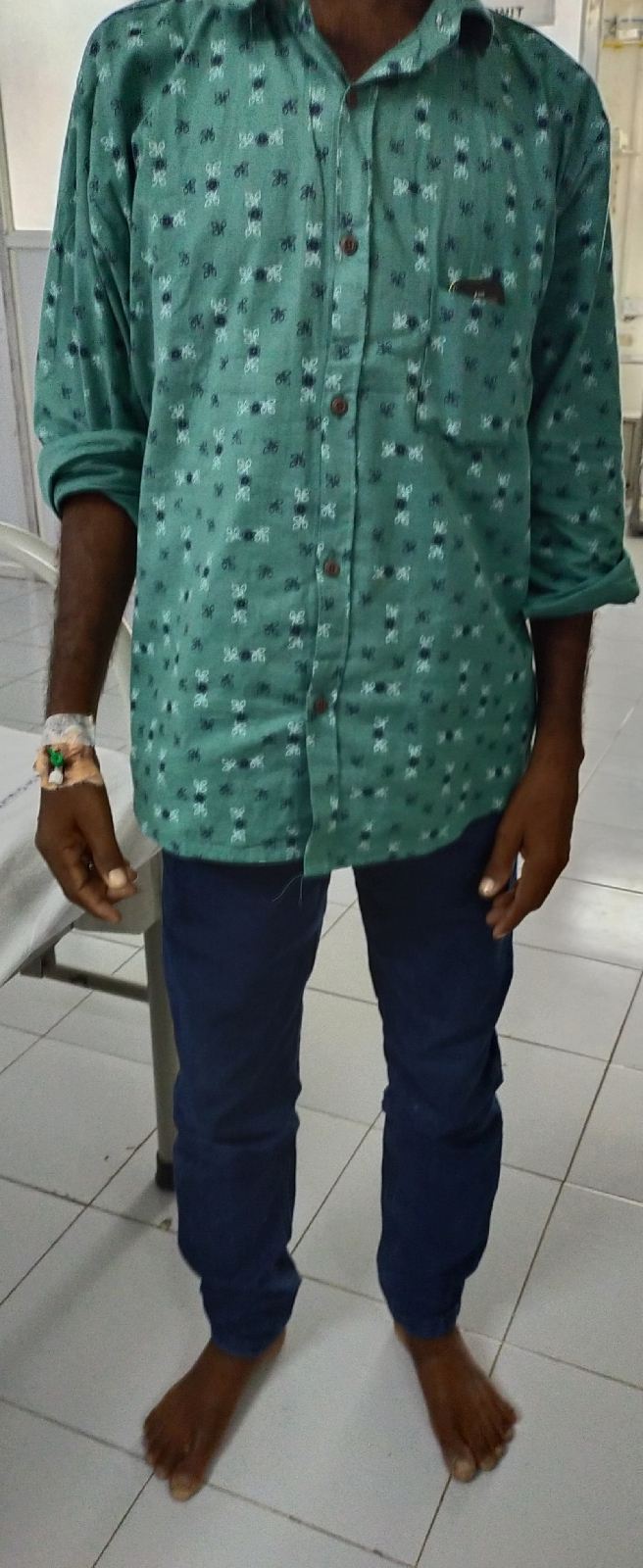
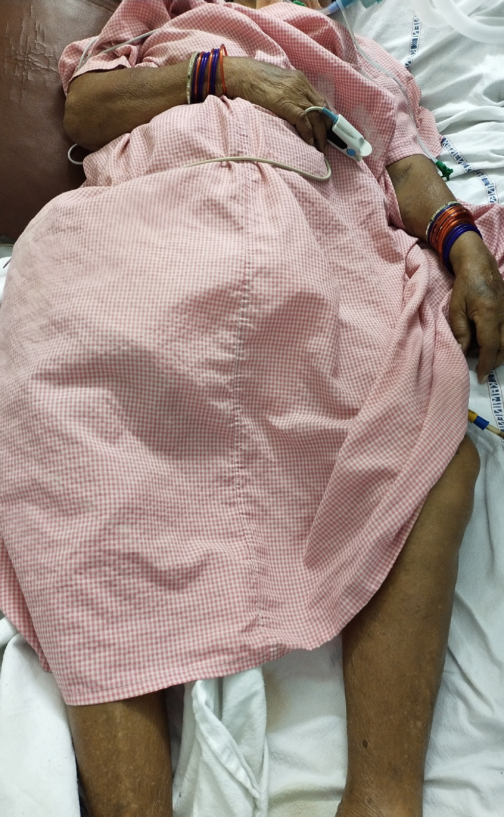
" CASE OF A 35 YEAR OLD MALE "
I have been given this case to solve in an attempt to understand the topic of "patient clinical data analysis" to develop my competency in reading and comprehending clinical data including history, clinical findings, investigations and come up with a diagnosis and treatment plan.
YOU CAN FIND THE ORIGINAL CASE IN THE LINK GIVEN BELOW :
CHIEF COMPLAINTS :
- Fever - 1 month ago
- Shortness of Breath since 2 weeks
- Bilateral Pedal edema since 2 weeks
1)FEVER :
- Onset - sudden
- duration - 1 month ago
- progression - gradually progressive
Given ANTIMALARIAL DRUGS as medication by RMP
2)SOB :
- Onset- sudden
- Duration- 2 weeks
- Progression- Gradually progressive (NYHA class 3)
- Relieving factors- Medication (It was not completely relieved but decreased from class 3 to class 2)
May be a RESPIRATORY cause but since he has no other associated complaints like hemoptysis, cough ,cold,wheezing ( NOT SUGGESTIVE OF RESPIRATORY PATHOLOGY)
3).BILATERAL PEDAL EDEMA :
- Onset- sudden
- Duration- 2 weeks
- Site- Bilateral Pedal edema extending upto both the knees
- Type- Pitting
- Progression- Gradually progressive
ASSOCIATED WITH GENERALIZED WEAKNESS AND PND SINCE 1 MONTH
PAST HISTORY:
- No history of similar complaints in the past
- Not a known case of DM ,HTN,Epilepsy,CVA,CAD
FAMILY HISTORY :
No significant family history
According to the history , system to be closely examined is CVS
POSITIVE CLINICAL FINDINGS :
- BILATERAL PEDAL EDEMA
- RAISED JVP (20 cms)
- EARLY INSPIRATORY CREPTS ON RIGHT SIDE
-Pneumonia
-Heart failure
-Interstitial Lung Diseases (No data suggestive of that)
- RAISED JVP ALSO SUPPORTS HEART FAILURE
INVESTIGATIONS :
- CBP- High PPBS and high FBS(Suggestive of Type 2 DM)
Requires other Investigations such as HbA1C
2.ECG -
- Ventricular Premature Complexes
- Right Axis Deviation
- Right Bundle Branch Block (because of 2 R waves in V1)
Various possible exaplanations:
- Heart disease due to high BP in the lungs (Pulmonary Hypertension)
- COPD (Because he is a smoker, but there is no cough)
- Pulmonary embolism
- Cardiomyopathy
- Myocarditis
3. USG abdomen-
- Grade 1 Fatty liver
- Mild ascites
- Moderate Right sided pleural effusion
4. 2D ECHO-
- Reduced ejection fraction i.e. 27% (<40%)
- IVC dilated (2-3 cm), not collapsing
- Mild TR
- Severe MR
- Trivial AR
- All the chambers are dilated
- Global hypokinesia
- Severe LV dysfunction
- Mild PAHT (could be responsible for the Right Heart Failure)
- No MS/AS
- No PE/LV clot
The ECHO findings are suggestive of Dilated Cardiomyopathy. In this case, it could be caused by-
- Diabetes (high Post prandial Blood sugar)
- Alcohol abuse
- Infections- viral, bacterial,parasitic,fungal (past history suggestive of fever)
5)LIPID PROFILE :
Low HDL
High LDL
6) LOW CREATININE LEVEL
( SUGGESTIVE OF CVS PATHOLOGY SUPPORTING HEART FAILURE )
The above features are suggestive of Dilated cardiomyopathy causing heart failure.. The possible causes for which in this case could be Myocarditis or Diabetes
Reason for Myocarditis in this patient could be -
- Viral infection (most common cause)
- Alcohol can directly injure the myocardium
- Could be from the anti-malarial medication (assuming it is Chloroquine/Hydroxychloroquine)
So in order to confirm the cause, Additional investigations required are
- ENDOMYCARDIAL BIOPSY - DIAGNOSTIC TOOL
- PCR - To know the causative virus (most commonly -Coxsackievirus group B, Parvovirus B19, HHV-6)
- CHEST X RAY
Anatomical location of the root cause is in the myocardium.
ETIOLOGY :
Pathophysiology which could explain the case:
TREATMENT :
*MEDICAL -
- ARB'S
- DIURETICS
- ACE INHIBITORS
- DIURETICS
- BLOOD THINNERS - To prevent clots
*SURGICAL -
- Cardiac resynchronization by biventricular pacemaker.
- Implantable cardioverter defibrillators (ICD).
- Conventional Surgeries for coronary artery disease
- Heart transplant
*LIFE STYLE CHANGES -
- Avoid alcohol
- Genetic counselling
- Proper diet -restriction od sodium
- Aerobic exercise
Things which I did not understand in this case are:
- Why was Anti-malarial drug prescribed ?
- What were his auscultatory findings?
- What is his occupation?
- Can antimalarial drug prescribed for few days lead to DCM?
- Murmurs?
References:
- Harrison's principles of internal medicine
- Davidson's principles and practice of medicine
- https://www.slideshare.net/1171097100/approach-to-patient-with-dilated-cardiomyopathy?qid=7ab6dad0-53ba-4f97-b2ae-b1286ab7c0d0&v=&b=&from_search=5
- https://www.ncbi.nlm.nih.gov/pmc/articles/PMC5908263/#:~:text=The%20landmark%20study%20from%20Kuhl,failure%2C%20respectively%20%5B1%5D.
- https://books.google.co.in/books?id=b5wADkB9oDoC&pg=PA74&dq=dcm&hl=en&sa=X&ved=0ahUKEwjX2N_b497pAhVIzDgGHRIpCFEQ6AEIXzAH#v=onepage&q=dcm&f=
- https://www.123sonography.com/ebook/echocardiographic-features-dilated-cardiomyopathy
- https://www.cedars-sinai.org/health-library/diseases-and-conditions/r/right-bundle-branch-block.html


